Alive and Alcohol Free logoAlive and
Alcohol Free

A visual journey

My Story in Pictures

From the depths of illness to the gift of a second chance — these images trace the road from survival to a life truly lived, alive and alcohol free.

The Liver Transplant

A liver transplant replaces a failing liver with a healthy donor liver. The images below explain what that operation involves.
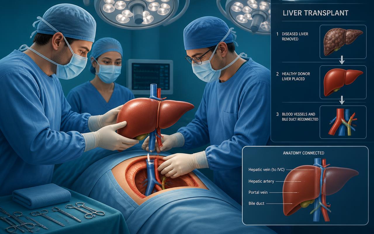
Medical illustration of a surgical team performing a liver transplant, with diagrams of the donor liver and reconnected blood vessels
Illustration of the transplant procedure: the diseased liver is removed, the donor liver is placed, and the blood vessels and bile duct are reconnected.
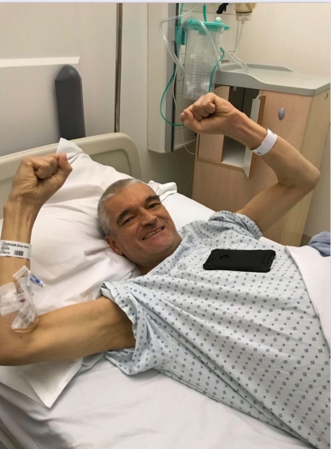
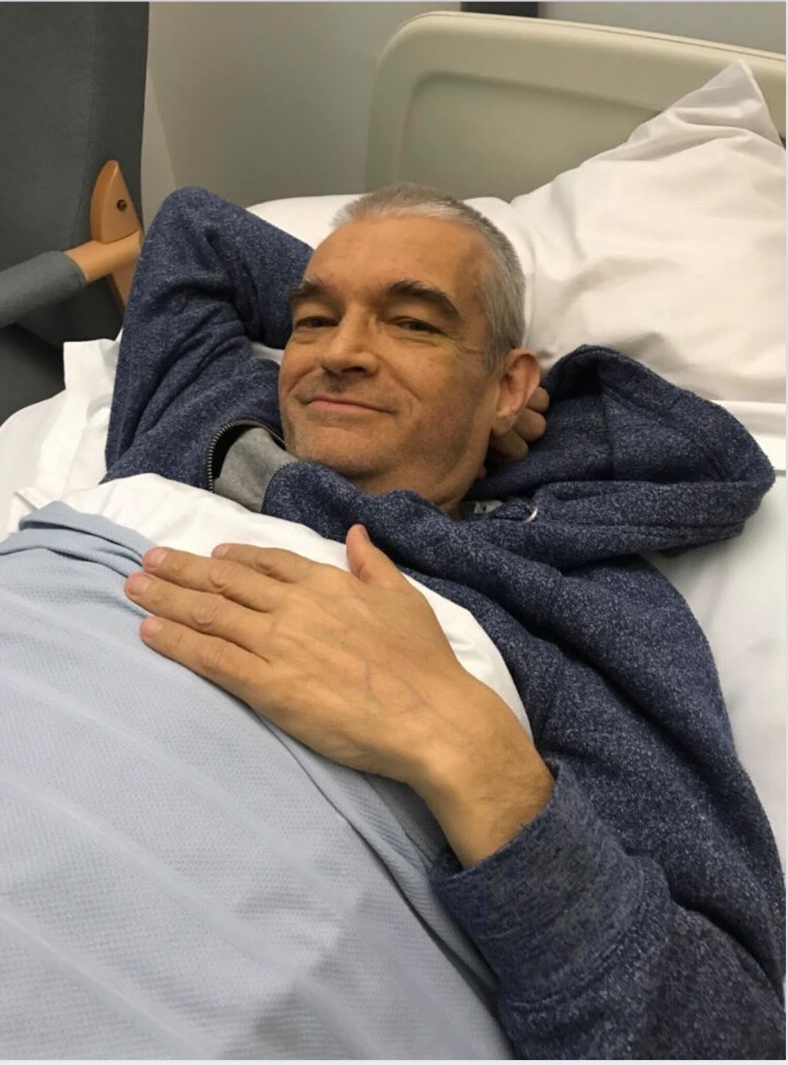
Brian resting in a hospital bed after his liver transplant
Transplant day! Just keeping warm before being taken down.
Brian celebrating in a hospital bed with both fists raised after his liver transplant
On the way down to theatre - celebrating a second chance at life.
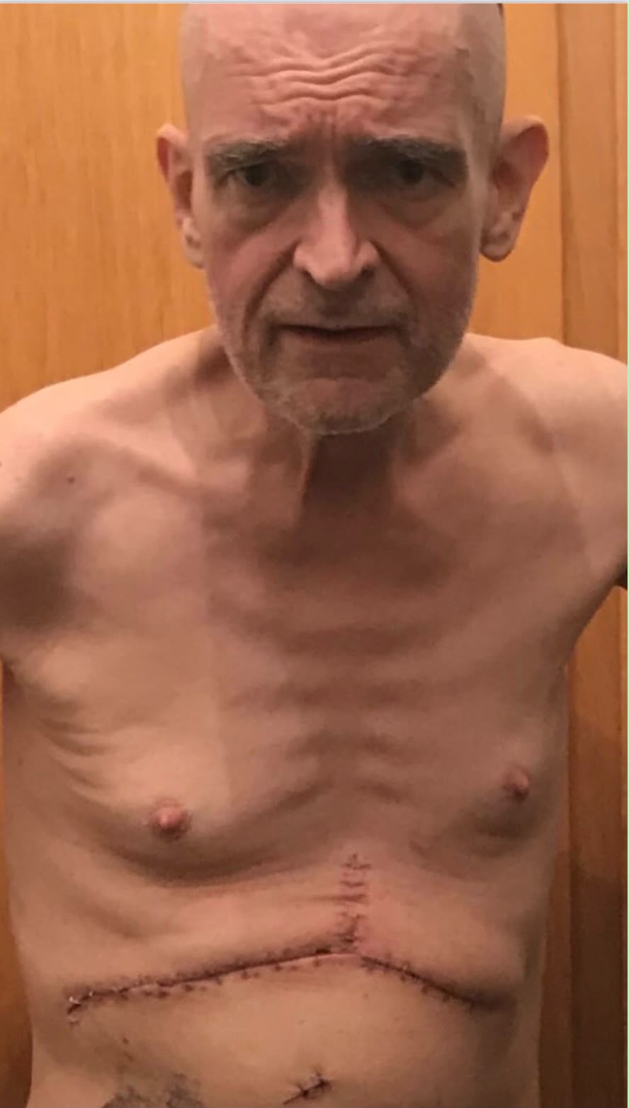
Brian showing the large Mercedes-shaped surgical scar across his abdomen after his liver transplant
Back at home and already feeling better even though I may not look so!!

ERCP and Biliary Stents

After my transplant, scar tissue restricted the flow of bile. I underwent six ERCP procedures to insert and remove stents that were meant to widen my bile ducts. Three separate attempts failed, and the stents could not keep the ducts open wide enough — that is when surgeons decided I needed the Roux-en-Y surgery to reconstruct my biliary system.
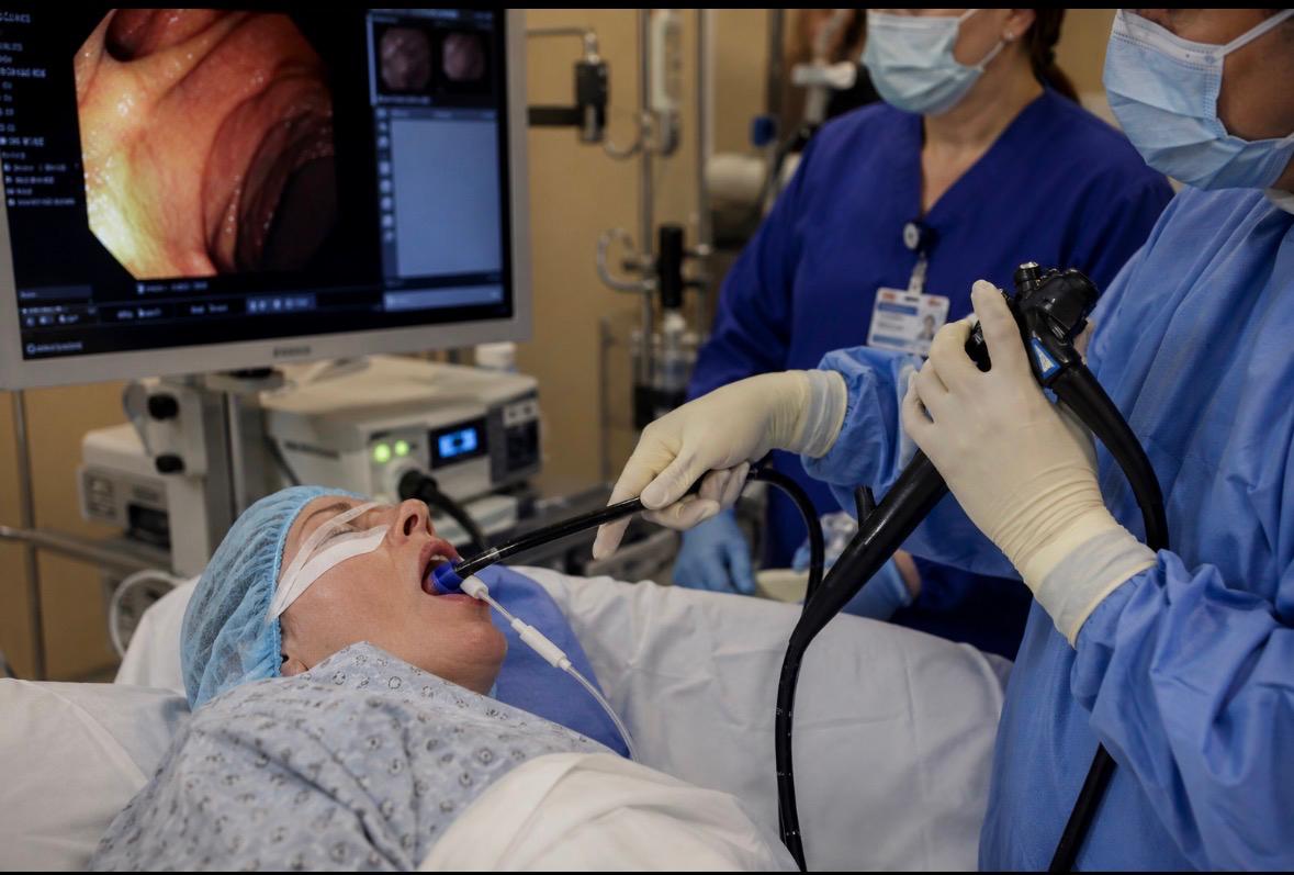
An ERCP procedure showing an endoscope passed through the mouth with the internal view visible on a monitor
ERCP — the procedure I had six times to try to keep my bile ducts open with stents.

Roux-en-Y Hepaticojejunostomy

After my transplant, scar tissue blocked the flow of bile. Surgeons rebuilt my biliary system, connecting my bile ducts to a loop of small intestine in a Y-shape so bile could flow freely again.
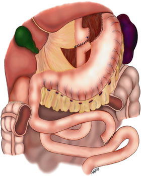
Schematic diagram of the Roux-en-Y anastomosis technique, showing the Y-shaped reconnection of the small bowel
A schematic of the Roux-en-Y technique — the same Y-shaped reconstruction used in my surgery. Image: Wikimedia Commons, by Topnife, licensed CC BY 2.5.
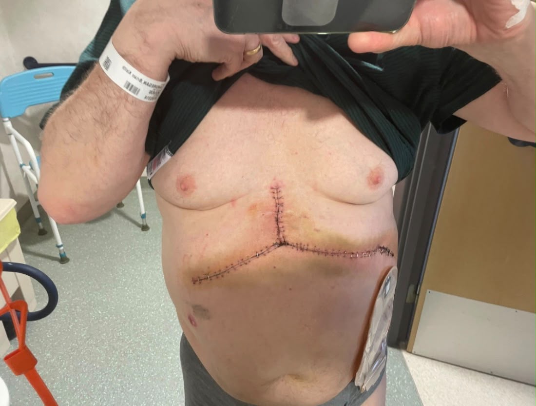
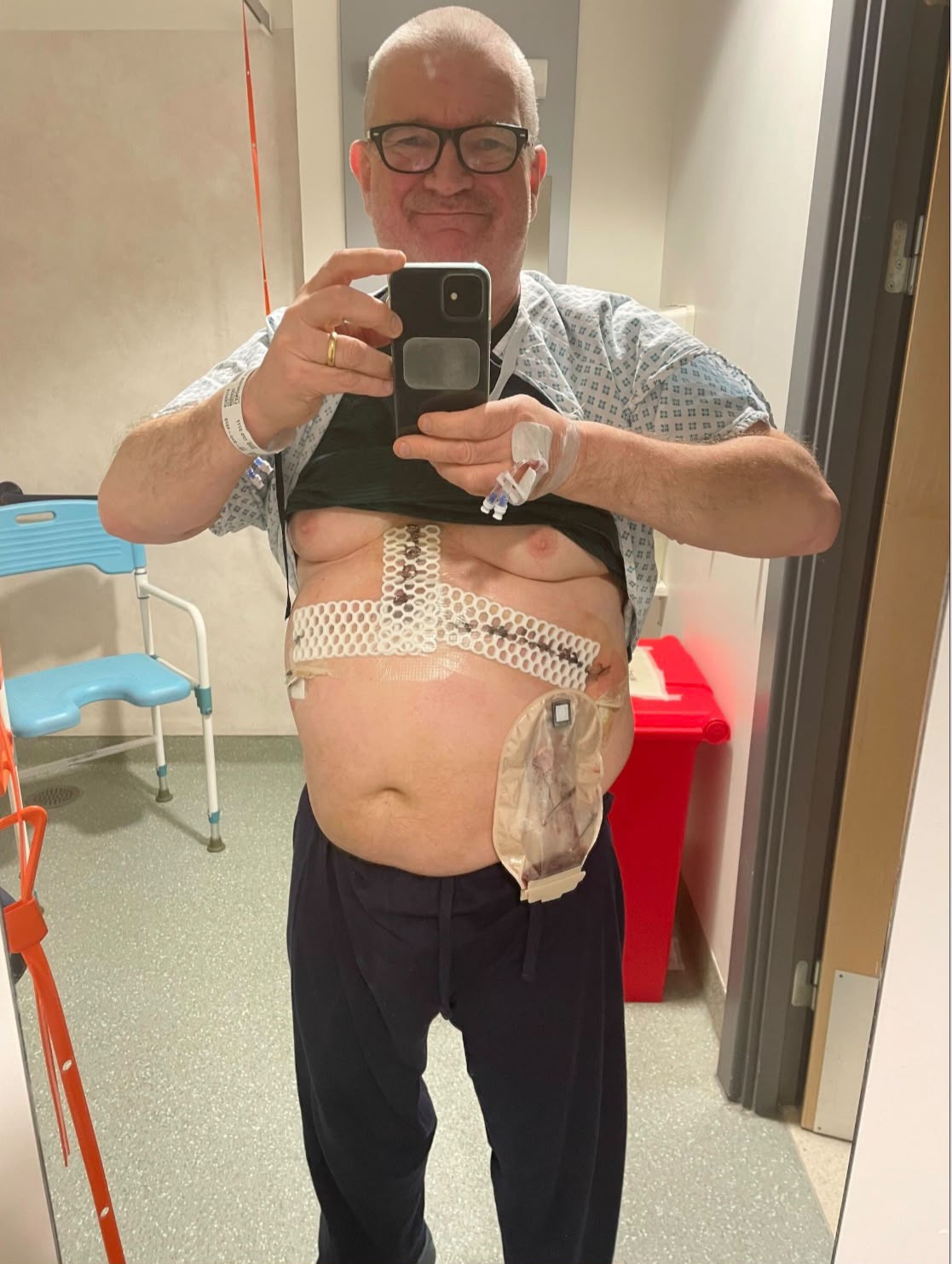
Recovering on the ward after Roux-en-Y surgery, with dressings and a drainage bag in place
Back on the ward after the Roux-en-Y surgery.
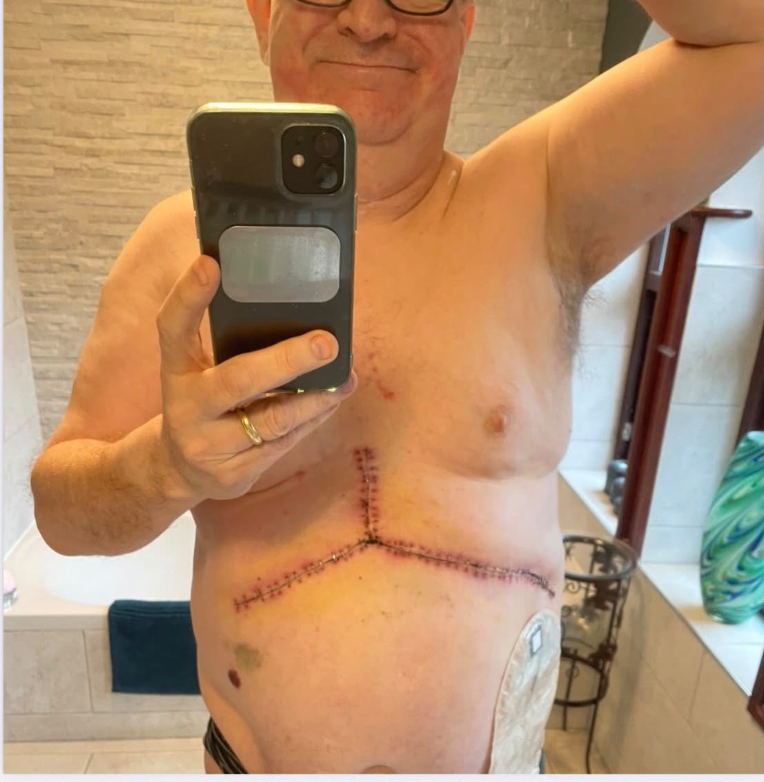
Healing Roux-en-Y surgical incision with clips still in place
The incision healing up, clips still in place.
At home after Roux-en-Y surgery, scar healing well
Home again and healing — getting stronger every day.

Ascites

Ascites is the build-up of fluid in the abdomen caused by advanced liver disease. It caused severe swelling, discomfort and breathlessness — I needed repeated drains to remove the fluid.
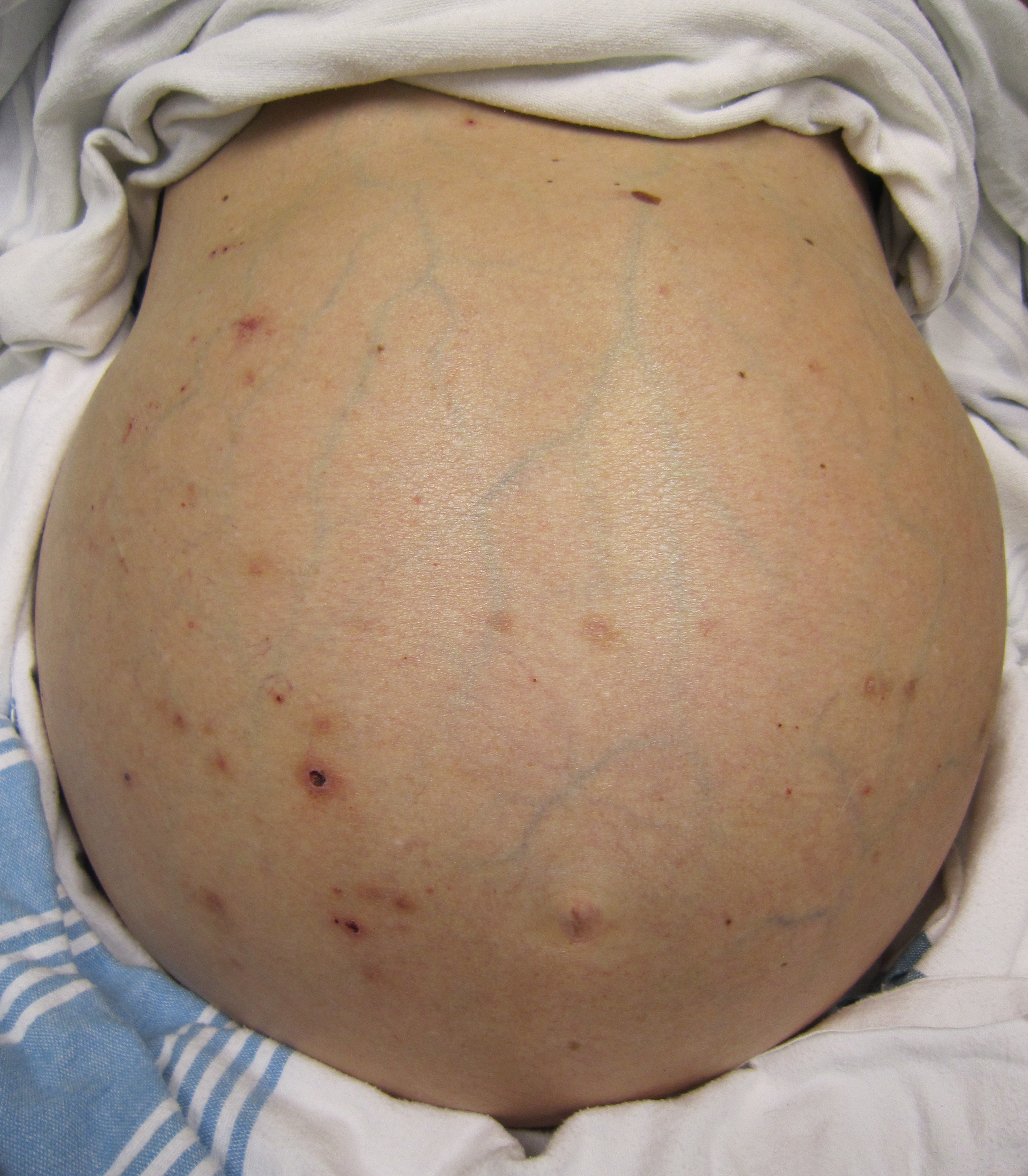
Clinical photograph of a man with a severely swollen abdomen caused by ascites from liver failure, with yellowing of the skin from jaundice
Ascites and jaundice caused by liver failure — the same signs my doctors recognised in me. Image: Wikimedia Commons, by James Heilman, MD, licensed CC BY-SA 3.0.

Abdominal Drains

To manage my ascites, I had three abdominal drains inserted to repeatedly remove the fluid that built up inside my abdomen — a procedure called paracentesis.
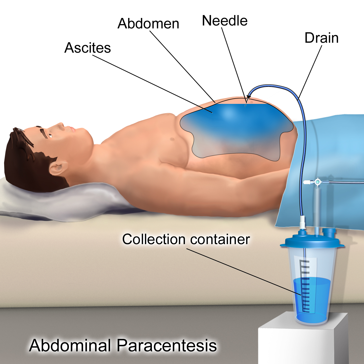
Medical illustration of abdominal paracentesis, showing a drain inserted through the abdominal wall to remove built-up fluid
How an abdominal drain works: a tube is passed through the abdominal wall so the built-up fluid can drain away safely. Image: Wikimedia Commons, by BruceBlaus / Blausen Medical, licensed CC BY 3.0.

Liver Biopsy

A liver biopsy takes a small sample of liver tissue so doctors can see exactly how much damage has been done — one of the many tests I went through on my journey.
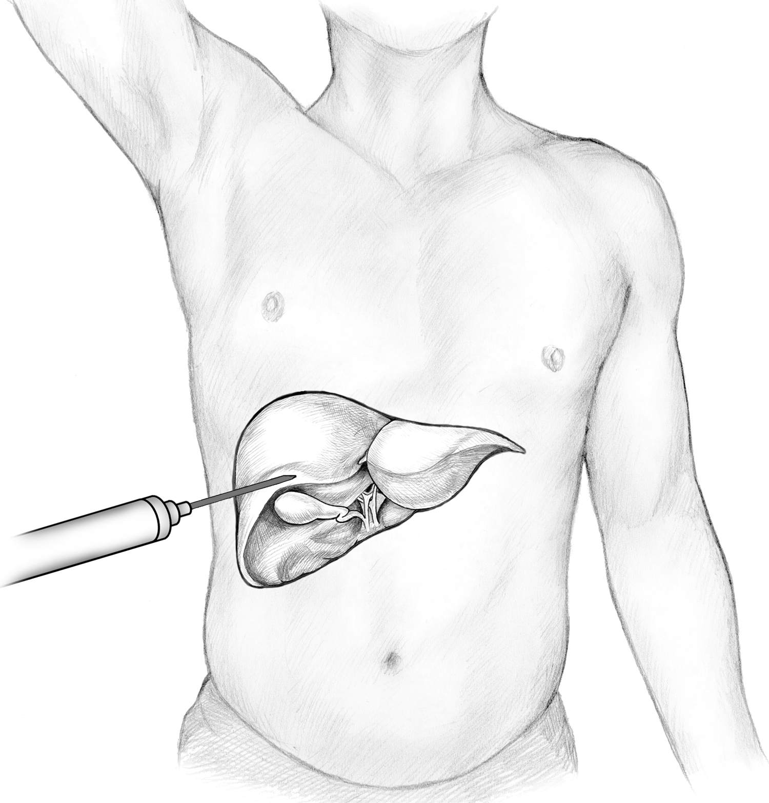
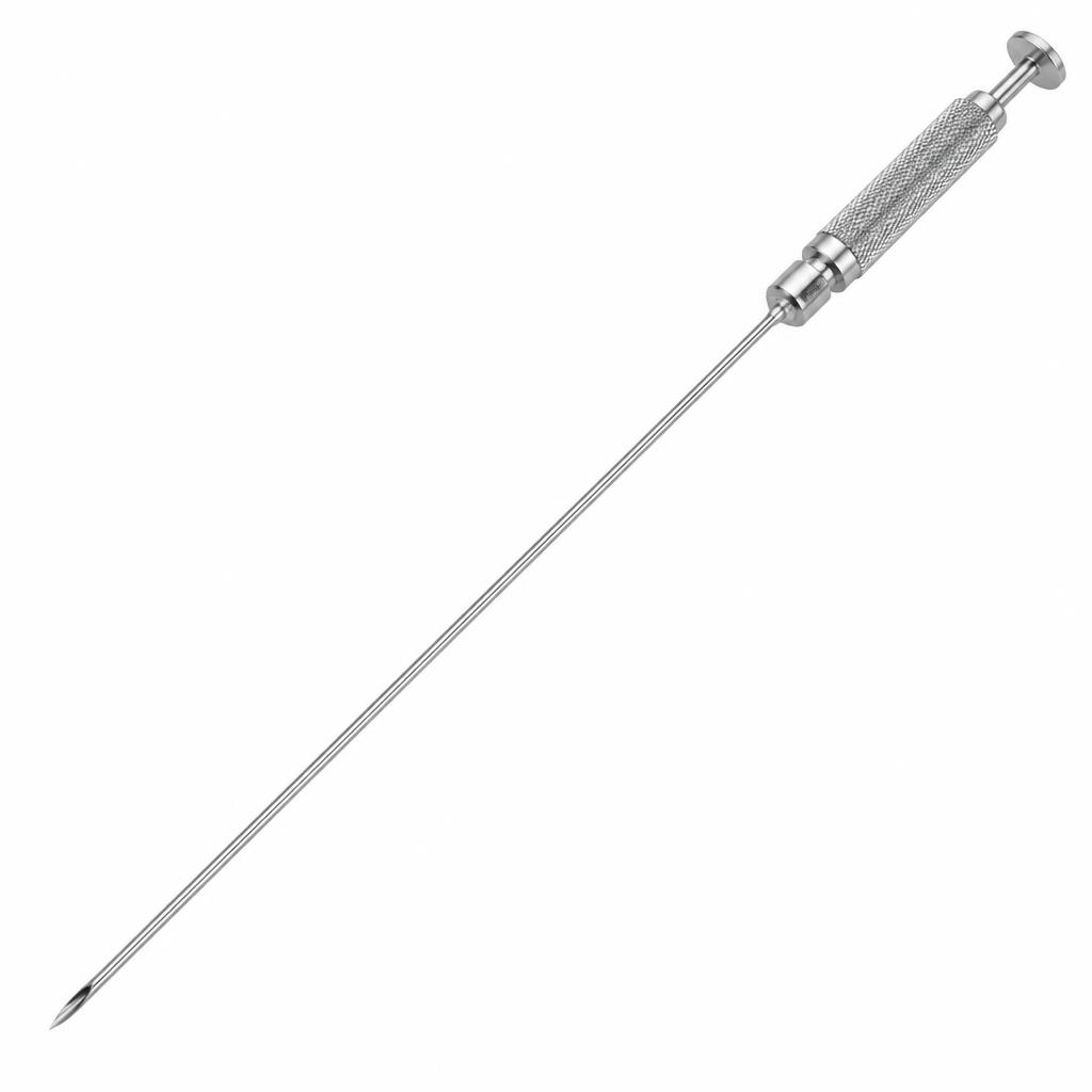
Illustration of a liver biopsy procedure, showing a needle inserted through the abdomen into the liver to take a tissue sample
How a liver biopsy is performed: a thin needle is passed through the skin into the liver to collect a tiny sample of tissue. Image: Wikimedia Commons, by NIDDK, public domain.
Long, slim hollow liver biopsy needle with a sharp bevel tip and handle grip
A long, fine liver biopsy needle — the kind used to pass through the skin and into the liver to collect a tissue sample, as was done during my biopsies.

IVC Filter Insertion

An inferior vena cava (IVC) filter is a small device placed in the large vein that carries blood back to the heart from the lower body. It catches blood clots before they can reach the lungs — another step in my medical care during this journey.
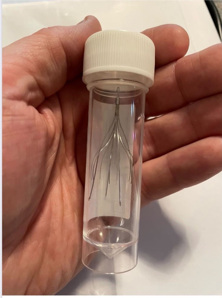
An IVC filter device held in a small clear vial, showing its wire legs designed to catch blood clots
The IVC filter — a tiny metal device that plays a big role in preventing clots from travelling to the lungs.

Nasogastric (NG) Feeding Tube

At my lowest weight and unable to eat safely, a feeding tube was passed through my nose, down my throat and into my stomach so I could receive nutrition and medication. It was uncomfortable, undignified and often made me gag — but it kept me alive.
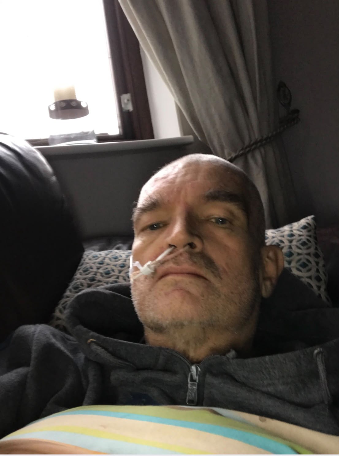
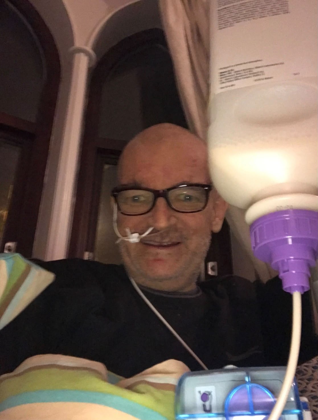
Brian lying back with a nasogastric feeding tube taped at his nose
The NG feeding tube in place — a thin line that carried the nutrition my body desperately needed.
Brian with the nasogastric feeding tube bottle in place for 12-hour feeds
Having me feeding bottle up my nose for 12 hours before having 12 hours without it. Not very comfortable

Insulin Pen Needles — Monthly Supplies

After my transplant, I developed secondary diabetes and now treat it as Type 1. I use around 150–180 needles every month to inject the insulin I need to keep my blood sugars at a safe level — a constant reminder of the routines that keep me alive.
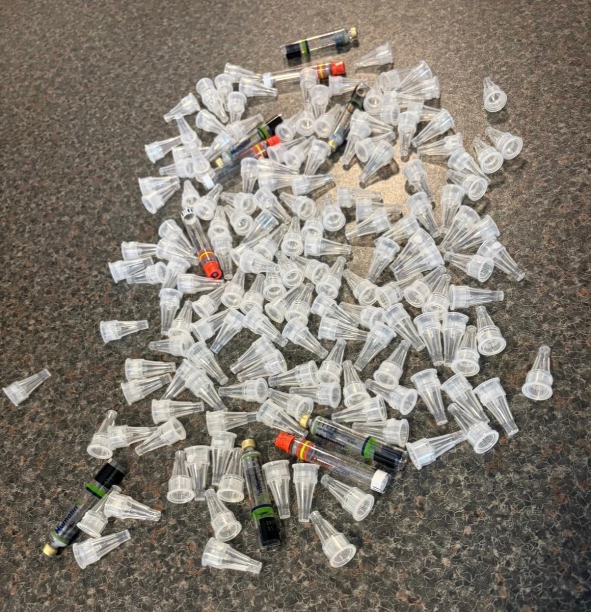
A large pile of used clear plastic insulin pen needle caps and syringes laid out on a worktop, representing one month's supply
One month's worth of insulin pen needles (approx. 150–180) — the daily routine that comes with secondary diabetes treated as Type 1.

Squamous Cell Carcinoma — Skin Cancer

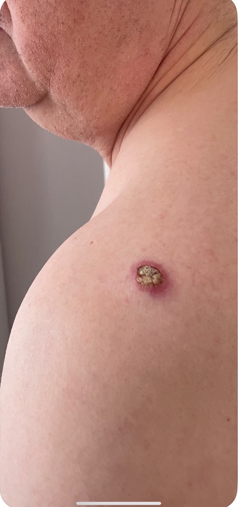
The immune suppression medication that I have to take for life has side effects, one of them being skin cancer. This squamous cell carcinoma had to be cut out TWICE — one more part of the price of a second chance.
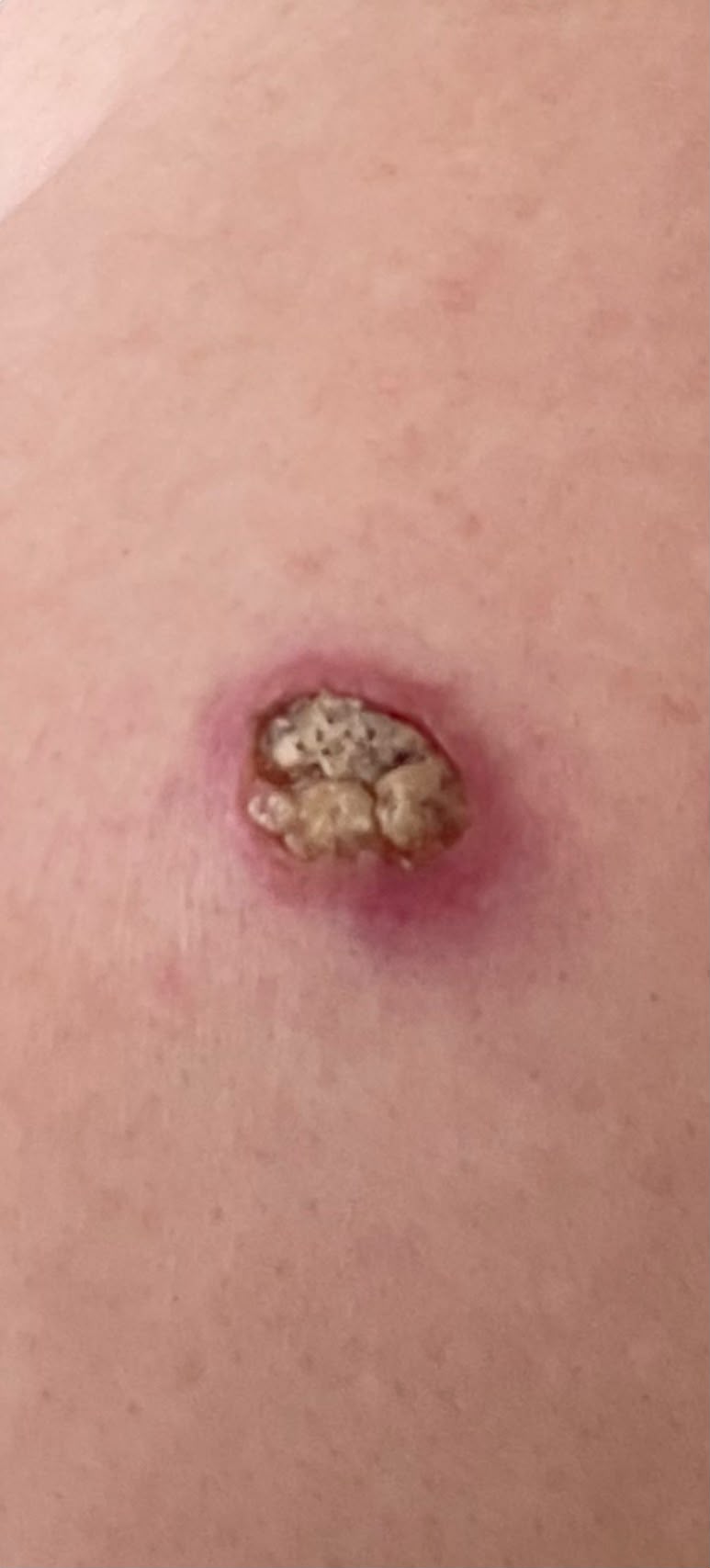
Close-up of a squamous cell carcinoma lesion on the skin
The squamous cell carcinoma close up — small, but not something to ignore.
Squamous cell carcinoma lesion on the shoulder
The same lesion on my shoulder, before it was removed.

Graphic content warning

This photograph shows surgery and an open wound. Some viewers may find it distressing.

This was the first operation to remove the cancer and its roots. I was told there was a chance the roots might not be fully removed, and that follow-up checks would confirm this. Sadly, some roots remained, so further surgery was needed.

Graphic content warning

This photograph shows surgery and an open wound. Some viewers may find it distressing.

This second surgical procedure removed the rest of the roots.